
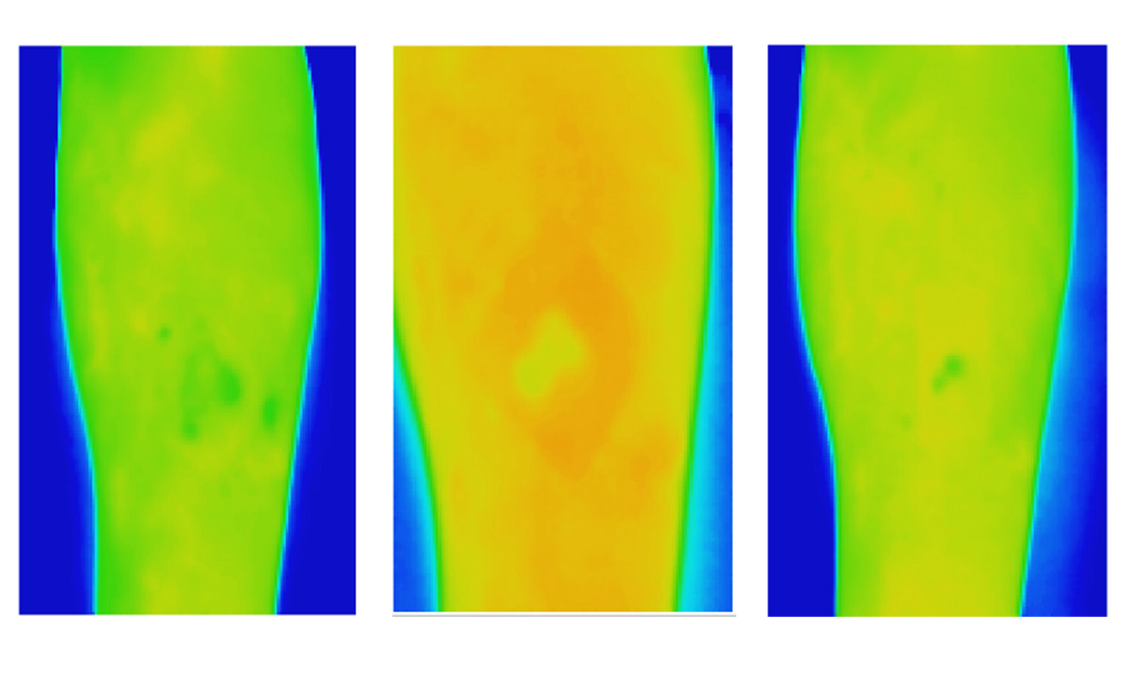
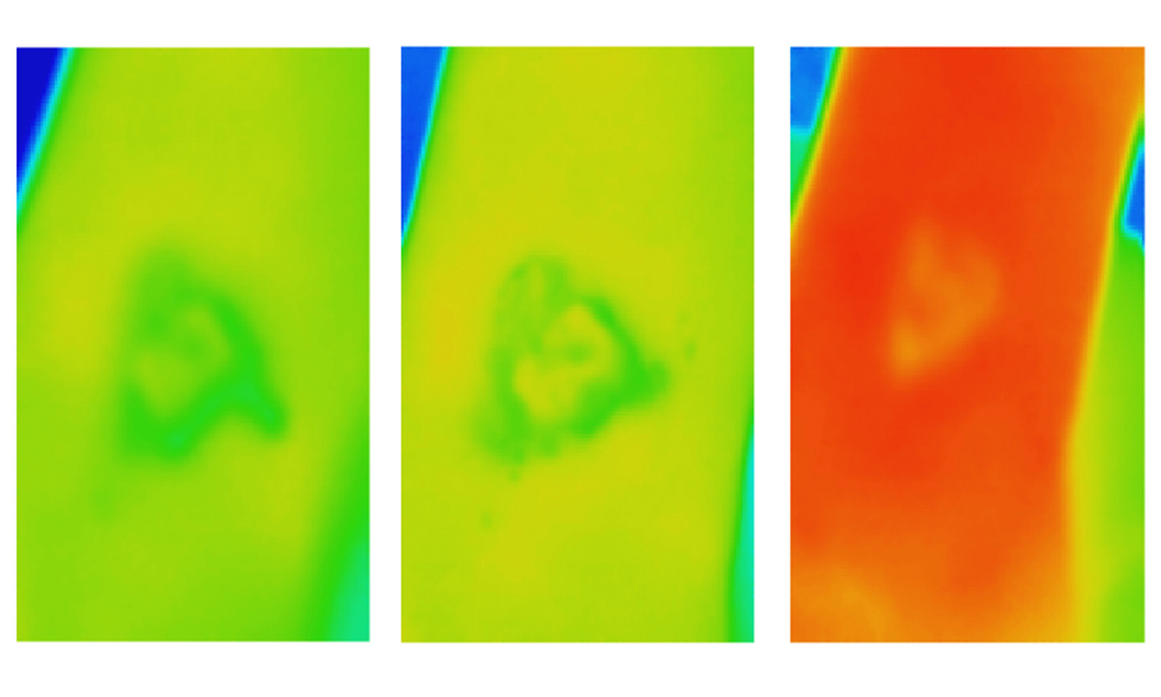
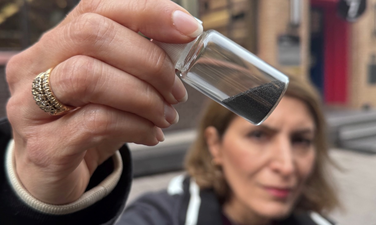
Magnetic invention removes ‘invisible’ microplastics plus some PFAS
RMIT University researchers have developed a water treatment material that rapidly removes micro and nano plastics and some PFAS, bringing the technology closer to real world use.
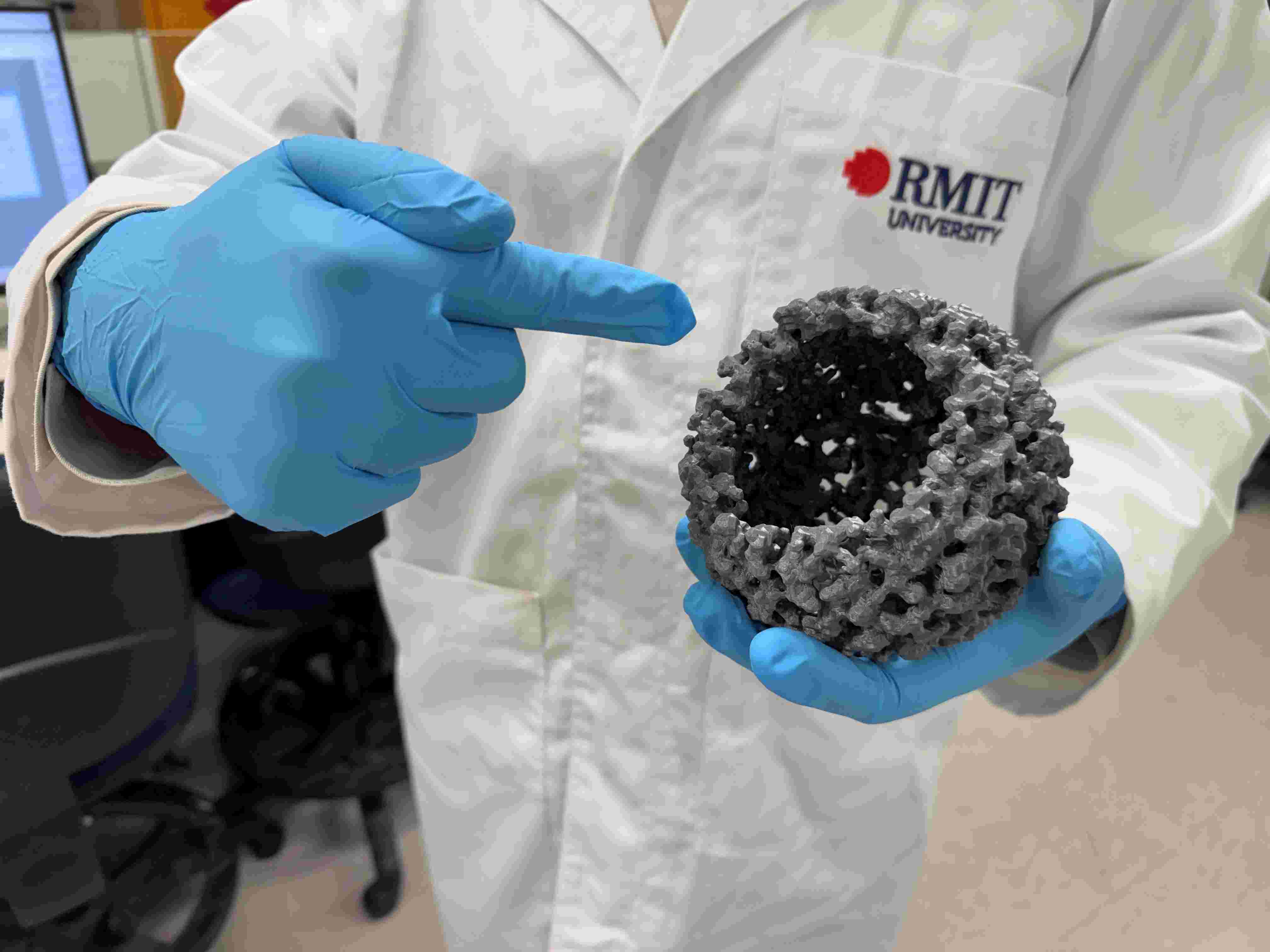
Low-cost innovation boosts green hydrogen production
RMIT researchers and international collaborators have demonstrated a new way to significantly increase green hydrogen production using low cost materials, offering a potential pathway to cheaper clean fuel.
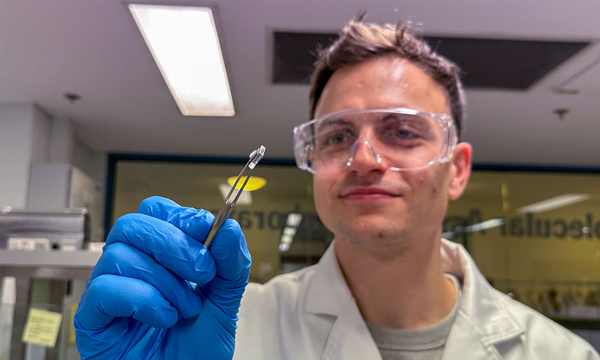
New study advances dry mRNA vaccine patch design
New research could help make future mRNA vaccines easier to store and distribute.
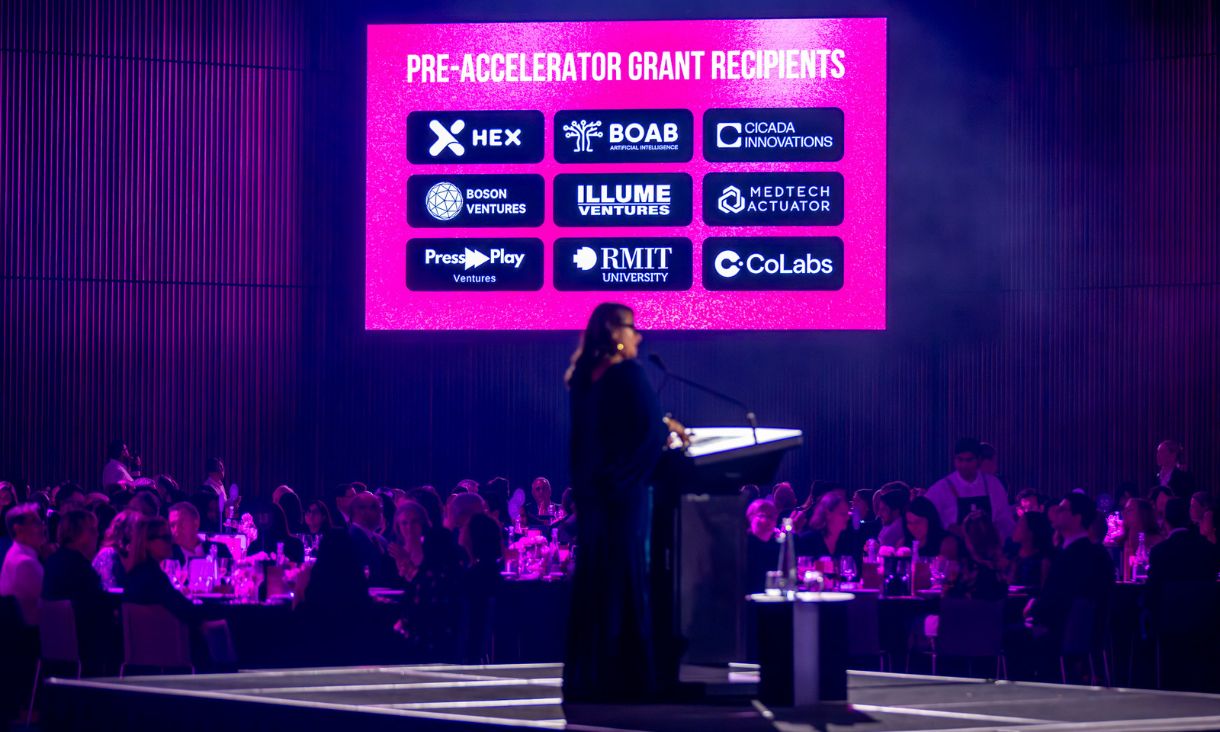
RMIT receives major grant to support AI and deeptech startups
RMIT has received about $400,000 in Victorian Government funding through Innovation Victoria to back early-career researchers as they test whether their AI, deeptech and MedTech ideas can become viable startups.