
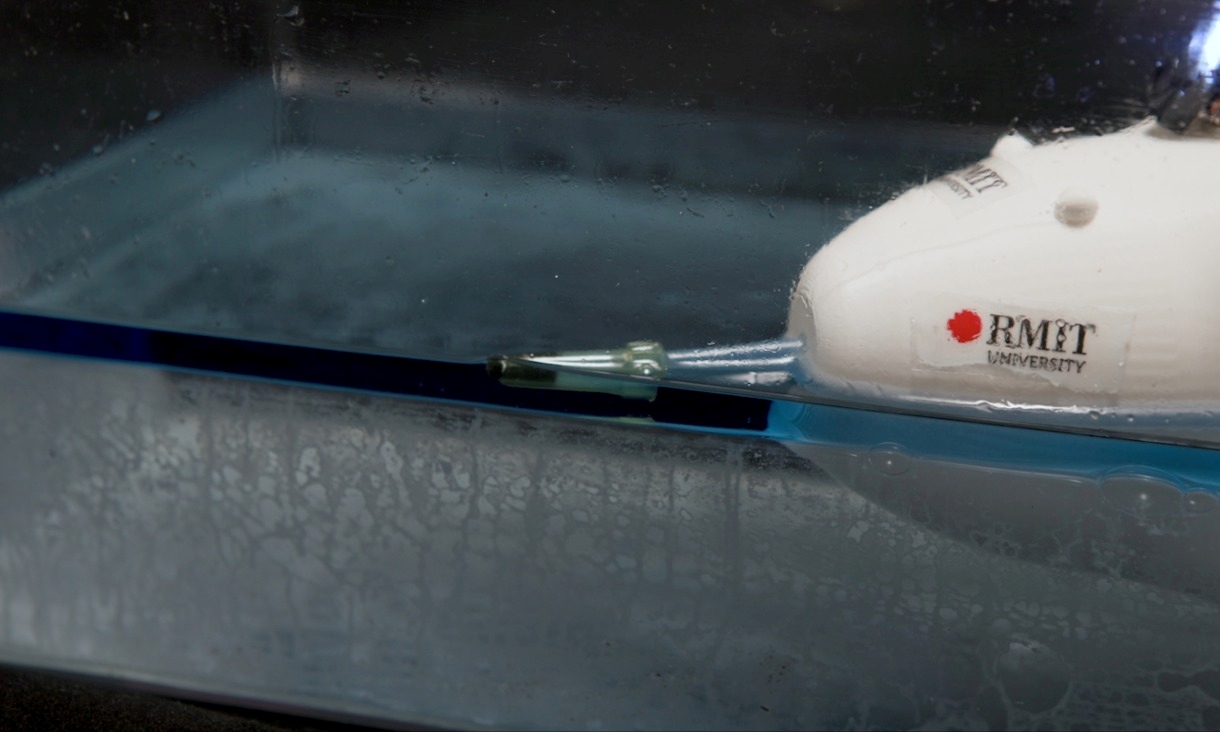
‘Sea creature’ minibot hoovers up oil spills
RMIT University engineers in Australia have built a remote-controlled minibot that hoovers up oil spills using an innovative filtering system inspired by sea urchins.
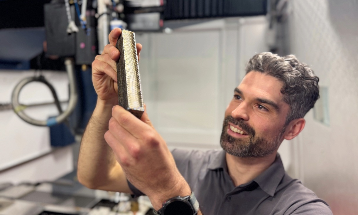
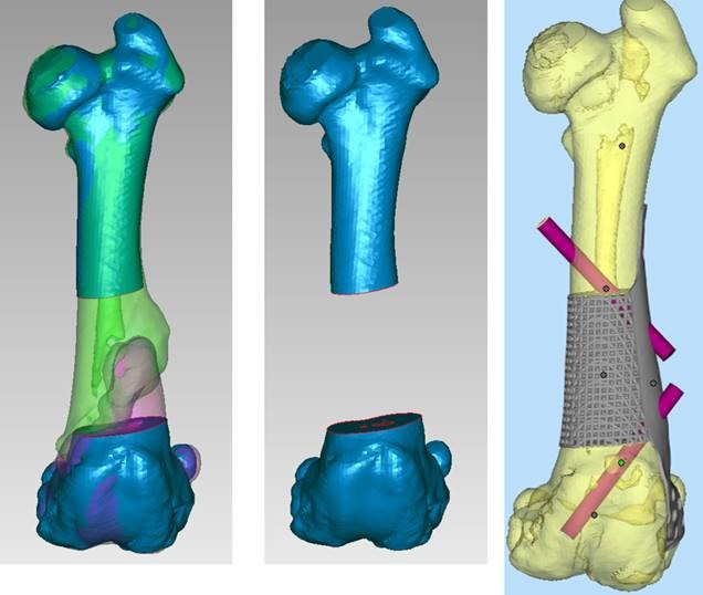
Stronger, cheaper titanium a 'leap forward' for industry
Engineers from RMIT University have produced a new type of 3D-printed titanium that’s about a third cheaper than commonly used titanium alloys.
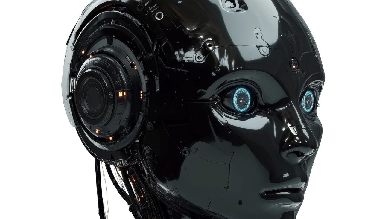
Tiny device promises new tech with a human touch
Engineers at RMIT University have invented a small ‘neuromorphic’ device that detects hand movement, stores memories and processes information like a human brain, without the need for an external computer.
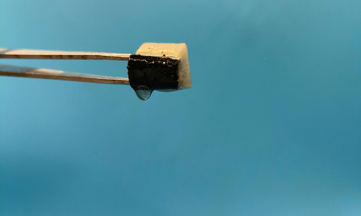
Smart spongy device captures water from thin air
Engineers from Australia and China have invented a sponge-like device that captures water from thin air and then releases it in a cup using the sun’s energy, even in low humidity where other technologies such as fog harvesting and radiative cooling have struggled.