Spare parts from small parts: novel scaffolds to grow muscle

Spare parts from small parts: novel scaffolds to grow muscle

RMIT biomedical engineers have successfully produced a 3D material that mimics nature to transform cells into muscle.

RMIT biomedical engineers have successfully produced a 3D material that mimics nature to transform cells into muscle.

The researchers have for the first time incorporated the natural processes of embryonic development to build a material that can more naturally communicate with stem cells for effective tissue repair.

The breakthrough offers hope to people suffering from injuries where their muscles cannot repair themselves due to trauma or disease, and our rapidly ageing population.

Led by Dr Richard Williams from RMIT University, the researchers have combined lab-made peptides with natural proteins and polymers to create a 3D “hydrogel scaffold”. 

The scaffold uses natural mechanisms to incorporate signals found in the natural developmental environment to support and engineer stem cells into muscle fibre.

“Our stem cells are at their best during development, where a scaffold drives them to produce all our tissues and organs; yet as people grow and age, they lose this ability and get a build-up of undesirable structures,” Williams said.

“So there is a real need for ways to repair and replace parts of the body as they wear out or become damaged, particularly as our population is now living longer.  

“Essentially, we make a scaffold that sends healthy signals to the cells to reorganise themselves to start to grow the small fibres that make up a healthy muscle.

“It’s like travelling back in time when our potential for tissue development and repair was at its best.”

Williams said it is a “simple process” to deliver complex structures inspired by the way nature pieces structures together during development, starting with only two cells.

“We have developed a simple, cheap, yet powerful toolkit that encourages different environments to let different tissues grow,” he said.

“The material properties of natural tissues, like muscles, are really complex and extremely difficult to mimic synthetically.

“Researchers have been producing ‘organising’ signals for years to understand how cells work but only in a flat, or 2D, environment.

“We have found a way to use the proteins present during embryonic development in a 3D scaffold, and use this extra dimension to potentially grow muscle.

“Our research shows that combining a hydrogel with these small proteins is easy and effective. In fact, just 24 to 72 hours after seeding the cells into the tissue, there was a noticeable reorganisation of individual cells into the multi-cellular structures that go on to make up functioning muscle.” 

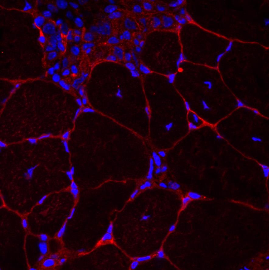
A detail of the muscle tissue the researchers are replicating with their novel 3D material. A detail of the muscle tissue the researchers are replicating with their novel 3D material.


The RMIT researchers are now exploring how they can bioprint muscle with the stem cells already embedded to put nerves, blood vessels and muscles together into lab grown “spare parts”.

The research has been carried out at RMIT University in Melbourne Australia, in collaboration with St Vincent’s Hospital Melbourne, Deakin University and the Australian National University. 

The scaffold has been developed in Melbourne’s BioFab3D@AMCD lab – Australia’s first robotics and biomedical engineering centre embedded within a hospital.

The researchers’ findings are published in Biomacromolecules.

How the 3D scaffold works

During embryonic development, stem cells exist within a specific and unique scaffold that allows them to organise into complex structures, such as tissues.

As we age the scaffold changes and these signals are less effective or even wrong, and prevent tissue repair from occurring as it should.

The researchers have been able to replicate the developmental process by producing a 3D material that sends the right signals to stem cells to help replace muscle fibres and repair damage.

The researchers’ method is based on a hydrogel scaffold – a delicate web formed from water and proteins. The scaffold is decorated with the proteins, and provides the structure and signals that the cells need to make muscles grow.

Using the simple, yet effective process, the researchers can take the signals and use them to tell the cells to behave in a certain way.

For interviews:
Dr Richard Williams, +
 61 3 9925 6642, +61 414 944 377 or richard.williams@rmit.edu.au


For general media enquiries:
James Giggacher, +61 3 9925 4143, +61 413 665 143 or james.giggacher@rmit.edu.au

Gosia Kaszubska, +61 3 9925 3176, +61 417 510 735 or gosia.kaszubska@rmit.edu.au

21 February 2018

Share

21 February 2018

Share

  • Science and technology

Related News

aboriginal flag
torres strait flag

Acknowledgement of Country

RMIT University acknowledges the people of the Woi wurrung and Boon wurrung language groups of the eastern Kulin Nation on whose unceded lands we conduct the business of the University. RMIT University respectfully acknowledges their Ancestors and Elders, past and present. RMIT also acknowledges the Traditional Custodians and their Ancestors of the lands and waters across Australia where we conduct our business - Artwork 'Sentient' by Hollie Johnson, Gunaikurnai and Monero Ngarigo.