Daniel Oldfield is a PhD candidate in the School of Science. He is passionate about the communication of science, and has found in microscopy an ideal way to engage public interest.
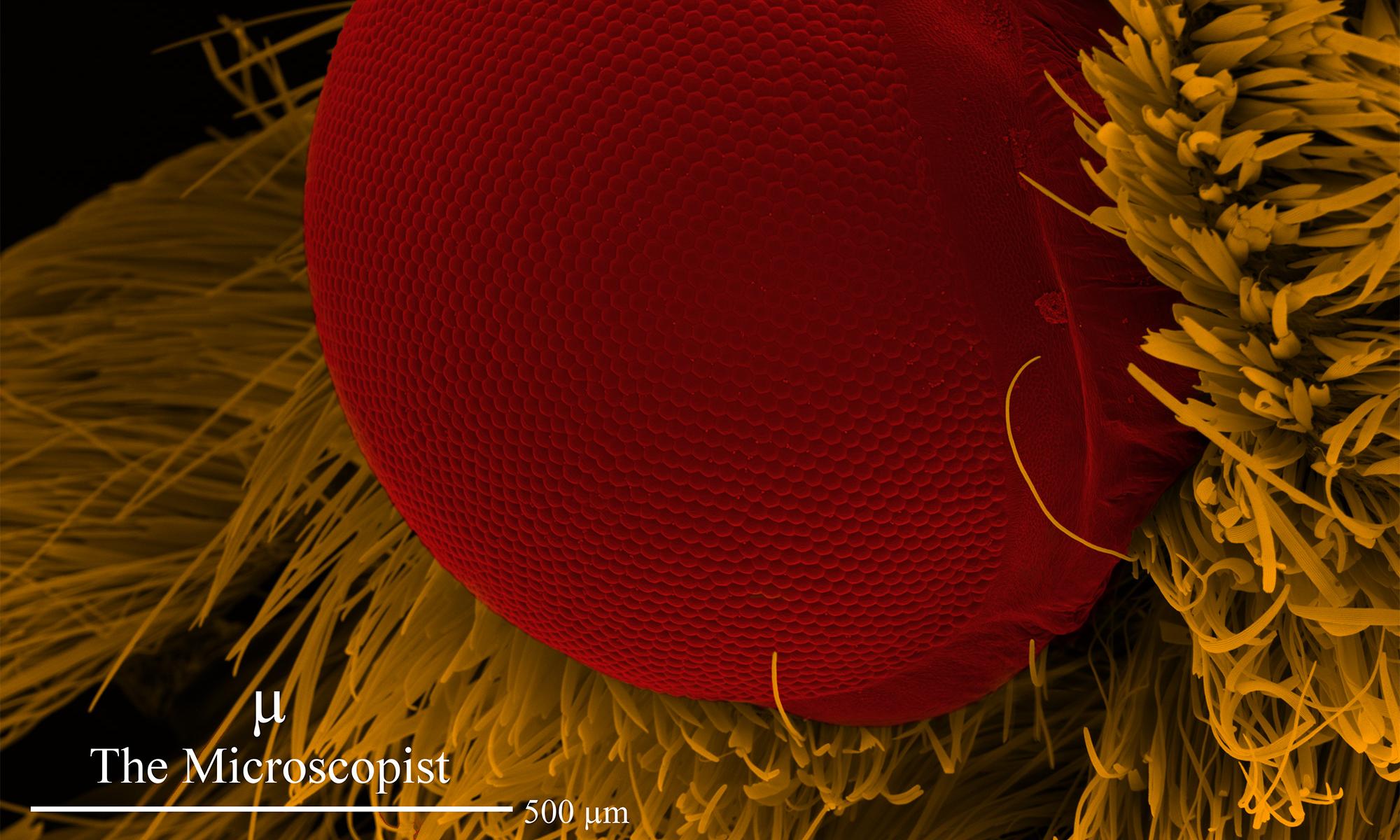 False colour image of a moth's eye viewed through a scanning electron microscope. © Daniel Oldfield.
What is the focus of your research?
My research focuses on the production of graphene.
Graphene has remarkable properties such as a high electron mobility and transparency. These properties give it potential for a range of electronic applications such as organic solar cells, ultra capacitors, biosensors, nano-electronics and batteries.
For large-scale graphene applications to be commercially viable, a fabrication method is needed that is controllable, scalable, cost effective and produces minimal defects.
A range of methods are currently used to produce graphene. Currently, chemical vapour deposition (CVD) is considered to produce the highest quality large-area sheets of graphene at the lowest cost.
However, CVD is very sensitive to growth conditions such as gas concentration, deposition time, temperature and substrate. Also, a transfer process is required post-deposition to place the graphene on a desirable substrate.
My research explores the use of physical vapour deposition (PVD) to resolve some of the issues surrounding the production of graphene.
How does the RMIT Microscopy and Microanalysis Facility support your work?
The RMIT Microscopy and Microanalysis Facility (RMMF) is a world class facility quipped with a variety of microscopes.
I use several instruments in the facility to characterise the graphene films that I grow using PVD.
Scanning electron microscopes work in a similar way to optical microscopes. Instead of using visible light and glass lenses, they use an electron beam and lenses created by magnetic fields.
The wavelength of an electron is much smaller than that of visible light. Hence a scanning electron microscope can resolve features which are less than 1 nm.
I assess the surface morphology and size of graphene domains using scanning electron microscopy.
Transmission electron microscopes generate a two dimensional image by sending an electron beam through a sample to create an image that is similar to a cross between a shadow and an x-ray scan.
For example, this technique allows scientists to see inside a cell to view all the individual components such as the cell membrane, vacuole, nucleus and mitochondria.
Transmission electron microscopy allows me to directly observe any disorder in the carbon atoms which are bonded together to form graphene.
Furthermore it enables me to see the separation between individual sheets of graphene, which is a distance of approximately 0.3 nm (that’s really small!).
An atomic force microscope can be used to create three dimensional images which reveal information about the height and roughness of a sample.
They create these images by tracing a fine needle across the surface of a sample in a similar way to how a blind person might read braille on a page of a book.
I used atomic force microscopy to measure the thickness of my graphene films.
What drew you to microscopy and what is it that continues to excite you about it?
Prior to commencing my PhD research I started working in the RMMF as a duty microscopist. As a microscopist my job involves looking after microscopy equipment and training facility clients.
Recently I have started a microscopy instagram account.
Through social media I hope to create a visual portfolio that not only showcases my ability as a microscopist, but provides a medium for other scientists to share their work.
It’s a real pleasure to share microscopy with the public.
Every time I look at a new sample I never know what to expect. Often I’m extremely surprised when I look at biological samples like insects and plants as they have some of the most interesting features.
In the future I hope to be able to represent science and contribute positively to science in a similar way to David Attenborough or Robyn Williams.
I believe the communication of science to the public can help improve decisions made by governments, corporations and communities, which leads to a higher quality of life for everyone.
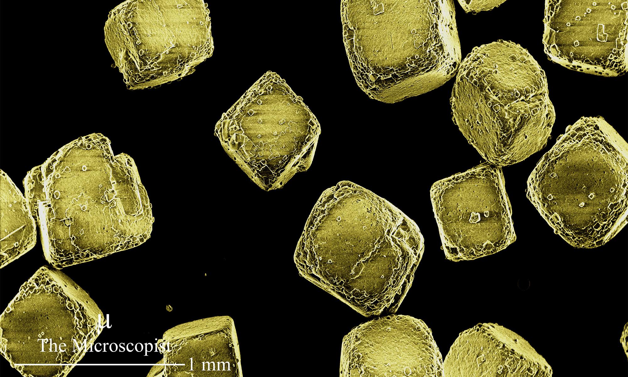 False colour image of salt crystals viewed through a scanning electron microscope. © Daniel Oldfield.