The following equipment and facilities are available to book by RMIT Researchers and members of Industry, Government Research Institutes, and external university researchers.
Transmission Electron Microscope (TEM)
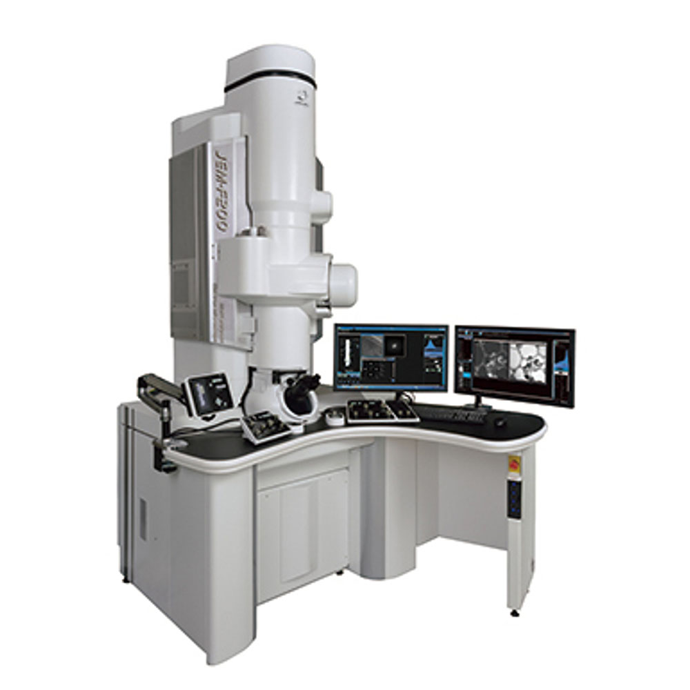
The JEOL JEM-F200 Transmission Electron Microscope (TEM) flagship is equipped with the state of the art Gatan Continuum GIF and EDS systems. The combination of the cold FEG JEM-F200 with a new generation of detector will allow high speed, high sensitivity EELS to be performed at high energy resolutions. In addition to conventional analysis, the RMMF has recently purchased a range of environmental TEM holders that will allow high sensitivity microanalysis to be performed on dynamic processes in-situ.
Some of its features include:
- Cold FEG with improved coherence over a Schottky FEG
- Improved lens and column stability
- sample auto loader (with compatible holders)
- TEM point resolution of
- STEM Point resolution of ** (BF) ** (DF)
- Gatan ContinuumS Spectrometer for super-fast EELS up to 0.45 eV energy resolution
- Duel EELS for simultaneous acquisition of two energy windows at different exposure
- Gatan Rio16 CMOS ( camera with image drift correction and rolling shutter 0.05 s exposure full frame
In-situ TEM sample holder suit for F200 and 2100F TEMs
- Protochips Fusion holder for in-situ electrical bias (up to 150 V) and heating (up to 1300C)
- Protochips Poseidon holder for in-situ liquid TEM including, heating (up to 100 C), in situ mixing of two fluids, and electro chemistry
- Gatan in-situ cooling (-170 C to -50 C) holder suitable for standard TEM specimens
- Gatan in-situ heating (RT to 800 C) holder with ±35 x-tilt suitable for standard TEM samples
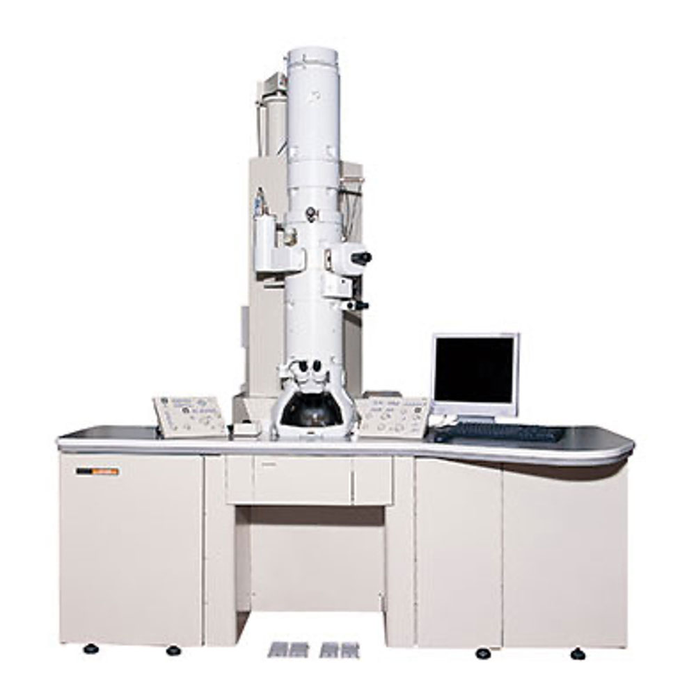
The JEOL 2100F transmission electron microscope (TEM) is a high resolution microscope capable of easily collecting lattice images, operating at either 80kV or 200kV. This machine is equipped with various spectrometers and can operate in scanning mode for precise control of the electron beam.
- Gatan OneView 4k Camera
- Tridium Gatan Imaging Filter (GIF) for performing EELS (typically 1.2 eV energy resoltuion)
- Oxford X-Maxn 80T EDS Detector mapping and drift correction.
- Gatan DigiScan System
- JEOL BF/DF STEM detectors
- Gatan BF/DF STEM detectors
- Single tilt holder with standard, Be (for EDS) and high tilt (±70 for tomography) quick change cartridges
- Double tilt holder ± 34 x and y tilt
- Rotation holder ± 34 tilt and rotation
In-situ TEM sample holder suit for F200 and 2100F TEMs
- Protochips Fusion holder for in-situ electrical bias (up to 150 V) and heating (up to 1300C)
- Protochips Poseidon holder for in-situ liquid TEM including, heating (up to 100 C), in situ mixing of two fluids, and electro chemistry
- Gatan in-situ cooling (-170 C to -50 C) holder suitable for standard TEM specimens
- Gatan in-situ heating (RT to 800 C) holder with ±35 x-tilt suitable for standard TEM samples
- Tungsten filament, operating at 100 kV or 80kV
- Gatan Orius SC600A CCD Camera (2kx2k)
- Rontec EDS detector
- Darkfield/brightfield
- Selected area electron diffraction (SAED)
- two position sample holder for rapid sample analysis
Scanning Electron Microscope (SEM)
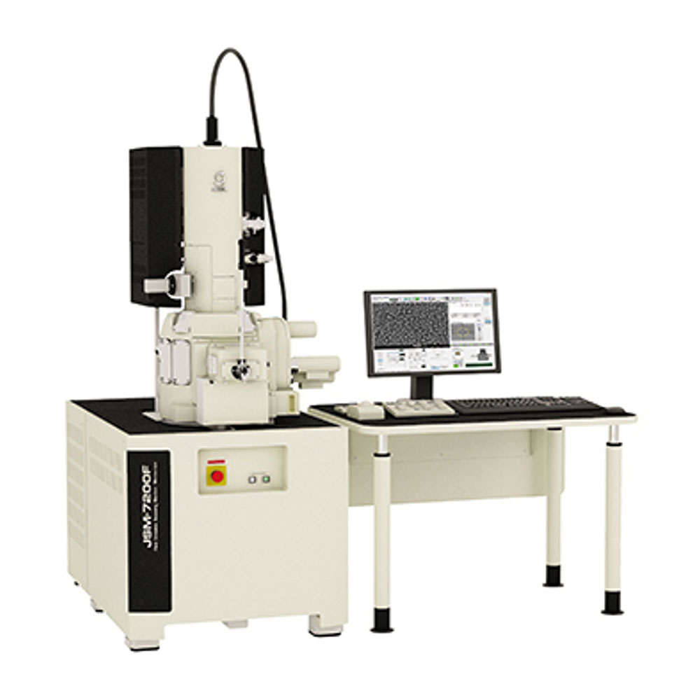
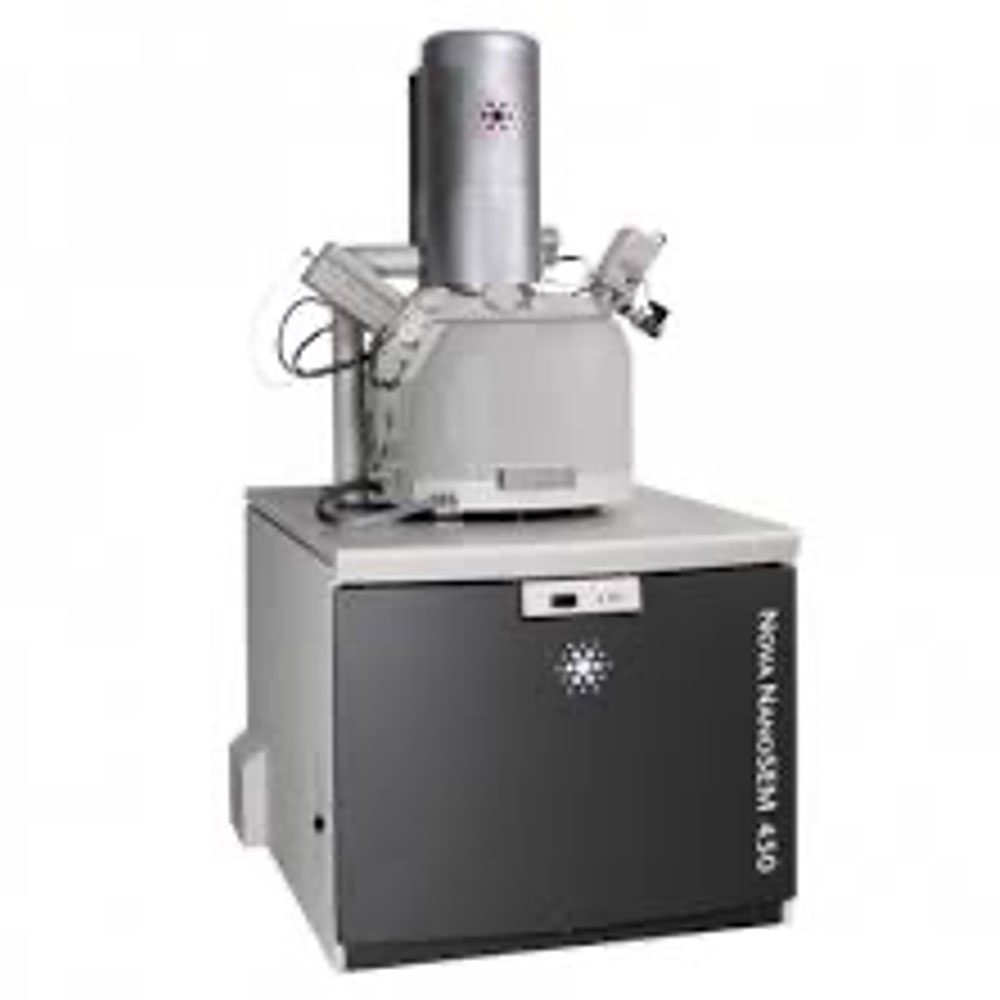
The JEOL JSM-7200F is equipped with a field emission gun (FEG) filament. Its primary purpose in the RMMF is to act as a high resolution and high throughput analytical SEM for EDS and EBSD.
It is capable of operating in a wide range of conditions, including:
Detectors
- Chamber mounted Everhart-Thornley (ETD) which can be operated in various modes.
- In Column Detector (UED)
- Retractable BSE detector
- Visible light camera for viewing sample/column
Additional Attachments
- Oxford XMax20 EDS Detector
- Oxford NordlysMax2 EBSD Detector
- Load lock to speed up sample loading and promote clean vacuum conditions.
- XXX Sample cleaner mounted to the load lock.
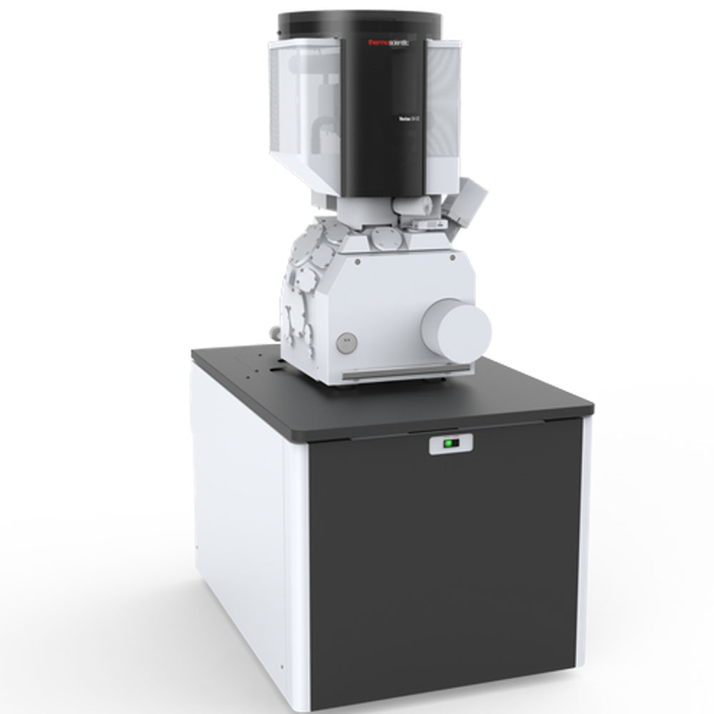
The FEI Verios is an ultra-high-resolution scanning electron microscope capable of resolving nanometre size features. Equipped with a number of in-lens and in-column detectors it is optimally suited to operating at low accelerating voltages which is beneficial for observing surface-based information or features from materials with a low atomic number.
Detectors
- Elstar in-lens SE detector (TLD-SE)
- Elstar in-lens BSE detector (TLD-BSE)
- Elstar in-column SE detector (ICD)
- Elstar in-column BSE detector (MD)
- Everhart-Thornley SE detector (ETD)
- IR camera for viewing sample/column
- Chamber mounted Nav-Cam+
- Retractable low voltage, high contrast solid-state backscatter electron detector (DBS)
- Integrated beam current measurement
Additional Attachments
- Oxford XMax30 EDS Detector
- Automatic Loadlock
Additional Software
- MAPS 2.0 (large area mapping) software
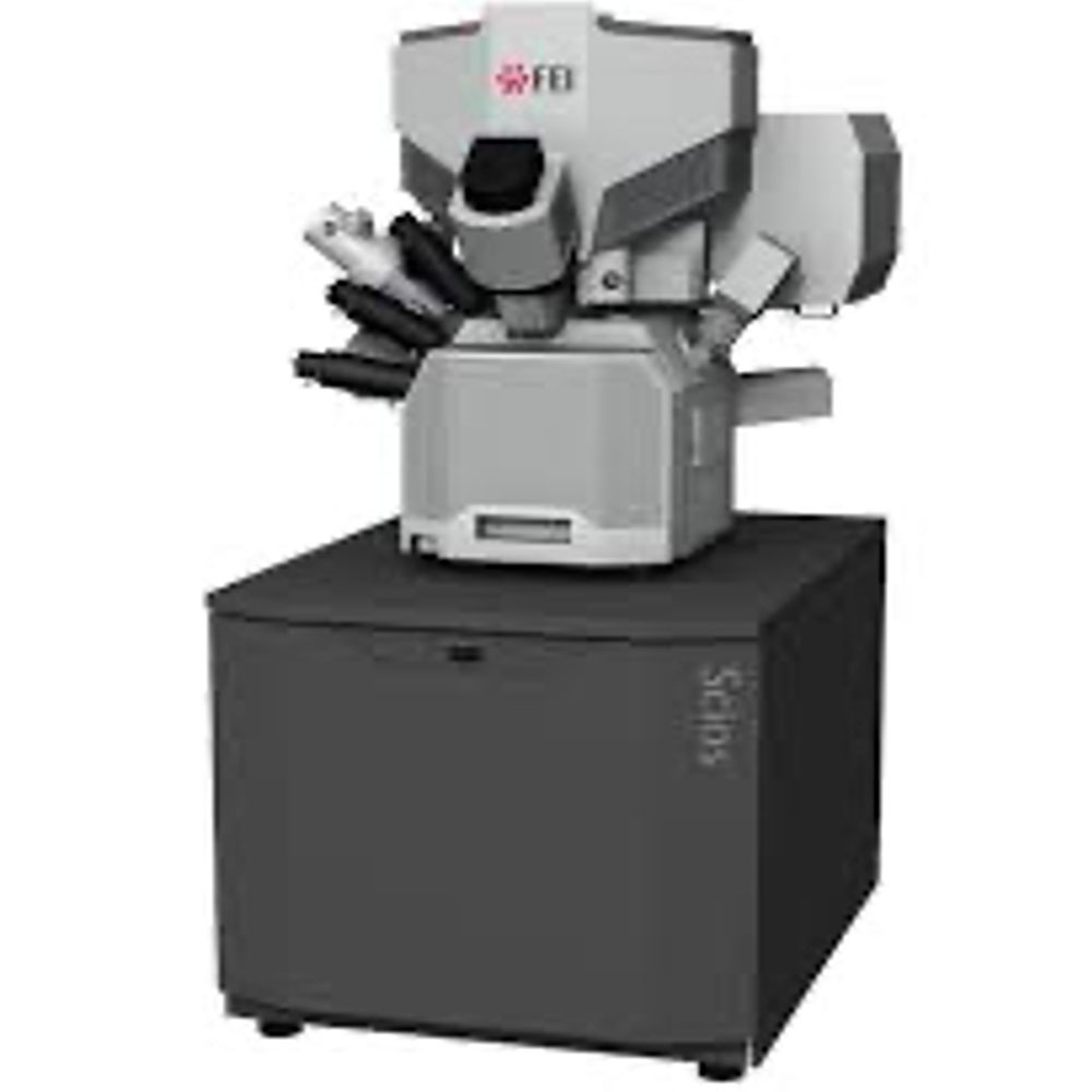
The Scios Ga ion column allows for accelerating voltages below 5kV, meaning that <5nm of damaged/implanted material is present on FIB lamella prepared for HRTEM. This, combined with the EZlift probe and STEM detector allow for extremely simple TEM lamella prep. The NiCol electron column allows for novel imaging modes (Optiplan, Optitilt and Analytical), which allows for enhanced contrast and resolution depending on the imaging required. This not only allows for excellent high-resolution imaging of magnetic samples, but also allows for improved contrast of biological samples when performing ‘Auto Slice and View’ to collect serial stack images for FIB-tomography. The large form chamber allows in situ experiments to easily take place, such as pico-indentation and heating using the Protochips Fusion SEM stage.
Detectors
- Up to four simultaneously detected signals
- Trinity Detection System (in-lens and in-column)
- T1 segmented lower in-lens detector
- T2 upper in-lens detector
- T3 retractable in-column detector
- Everhart-Thornley SED
- ICE detector (secondary electrons and ions)
- DBS retractable segmented under lens BSED
- STEM retractable segmented detector (BF, DF, HADF, HAADF)
- IR-CCD
- Nav-Cam chamber mounted camera
Additional Attachments
- Oxford XMax80 EDS Detector
- EZLift Micromanipulator Probe
- Platinum GIS
- Carbon GIS
Additional Software
- AutoTEM G2
- Auto Slice and View 4.0
- Auto FIB
The FEI Nova is an ultra high resolution (UHR) SEM equipped with a Field Emission Gun (FEG). With its combination of low vacuum mode and an ultra stable source, it makes the great candidate for analytic SEM needs. As such, as well as UHR imaging, it gets used a lot for:
- HR-SEM
- EDS mapping (elemental distribution)
- Electron-beam lithography (EBL)
The objective lens in the Nova can operate as either a standard pin-hole type lens, or as an immersion lens for UHR imaging. In immersion mode, the entire chamber acts as an objective lens, 'immersing' the sample in the magnetic field (no magnetic samples please!). This allows for sharper images and greater beam control at lower voltages.
Detectors
- Chamber mounted Everhart-Thornley (ETD) which can be operated in various modes.
- Through Lens Detector (TLD) which can be operated in a variety of modes.
- Polepiece mounted BSE detector
- Wide field low vacuum detector (LVD)
- Gaseous backscatter electron detector (GBED)
- Scanning transmission electron detector (STEM).
- IR camera for viewing sample/column
Additional Attachments
- Oxford XMax20 EDS Detector
- Nabity EBL Controller
- Port which can be used for Hysitron pico-indentation stage
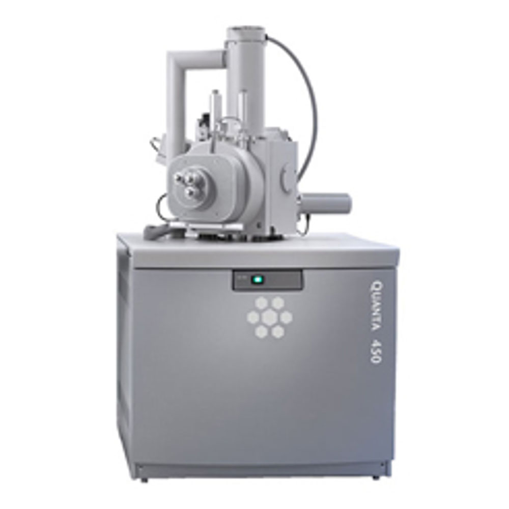
The Quanta can operate in either high vacuum or low vacuum mode, allowing a controllable humidity to exist in the chamber. The primary benefit of this is charge neutralisation, whereby it is possible to view insulating samples in the microscope without the need to coat them with a conductive gold film. This is useful if you have a sample which may react badly to the coating, or if a uniform coating isn't possible.
In addition to this, the Quanta can also operate in environmental (ESEM) mode. In this mode, the sample is chilled in a more humid environment than low vacuum mode, making it possible to image wet samples.
The FEI Quanta200 "Q1" is also equipped with the Gatan Alto Cryo stage (right hand side of image). This is a fully integrated cryo-transfer technique to rapidly freeze a sample, preserve its internal structure, then coat, fracture, and sublimate at temperatures less than -180 °C
Detectors
- Chamber mounted Everhart-Thornley (ETD) which can be operated in various modes.
- Large field low vacuum detector (LVD)
- Gaseous secondary electron detector (GSED) for environmental SEM mode.
- Polepiece mounted BSE detector
- IR camera for viewing sample/column
Additional Attachments on the "Q1"
- Oxford XMax20 EDS Detector
- Gatan Alto Cryo-Stage
Additional Attachments on the "Q2"
- Oxford EDS Detector (coming soon)
Modes/Test Conditions
- High Vacuum (Hi Vac)
- Low Vacuum (Low Vac)
- Environmental SEM (ESEM)
- Energy Dispersive X-ray
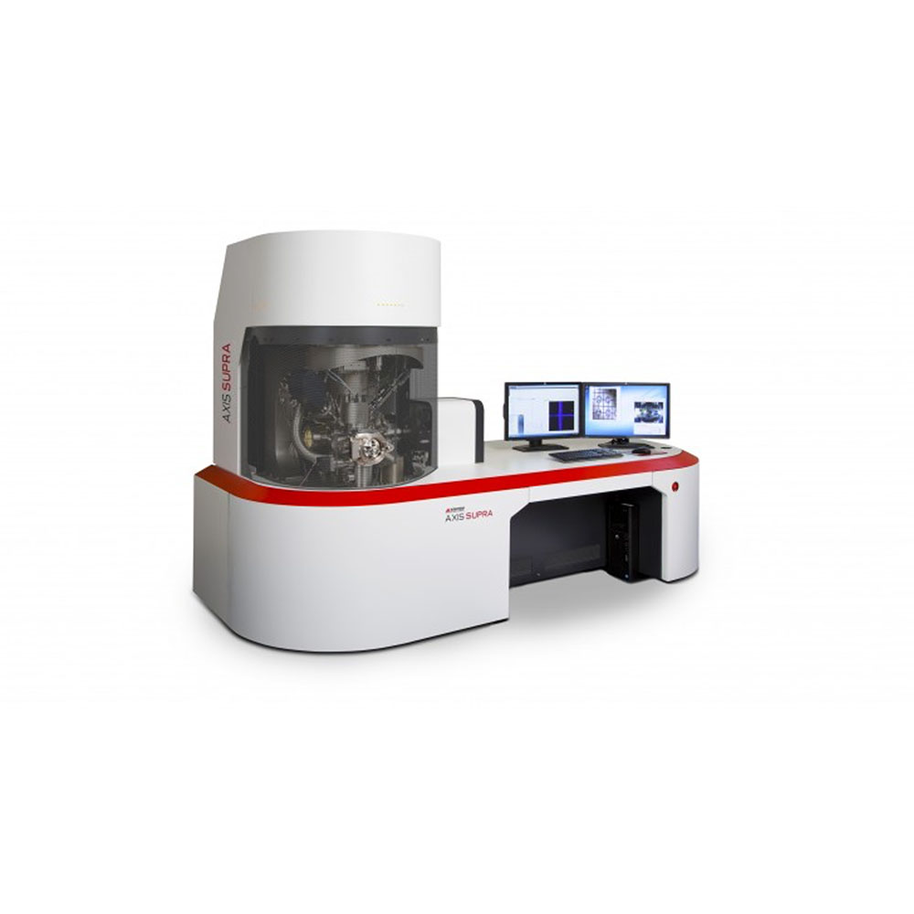
Surface Science
Photon sources
- Monochromated XPS - Aluminium Kα (1486.7 eV) or Silver La (2984.2 eV)
- UPS – He I (21.2 eV) or He II (40.8 eV)
- Analysis spot size – controlled by the collimation apertures. Spot diameter ranges from ~500 mm (Slot) to 15 mm.
Vacuum chambers
- The vacuum in the sample analysis chamber (SAC) can achieve as low as 1 x 10⁻⁹ Torr with several hours pumping time assuming a non- or minimally outgassing sample.
- The system is equipped with a flexilock that can achieve pressures as low as 1 x 10⁻⁸ Torr.
Additional Guns
- Co-axial charge neutraliser - The Supra is equipped with the patented AXIS charge neutralisation system that provides low energy thermionic electrons for charge compensation.
- Ion Gun (Minibeam 6) - Etching can be performed using the Minibeam 6 ion gun in either monoatomic or cluster modes.
Additional Features
- Heating/cooling - Temperature range ( -90°C – 1000°C)
- Catalysis cell for high pressure (up to 10 bar)/temperature gas reactions
- Ion scattering spectroscopy
- Stage tilting/Azimuthal holder - for ARXPS & rotational depth profiles
- XPS Imaging – spectromicroscopy (1µm resolution) using spherical mirror analyser and delay-line detector
- Air sensitive transporter & vacuum fracture stage
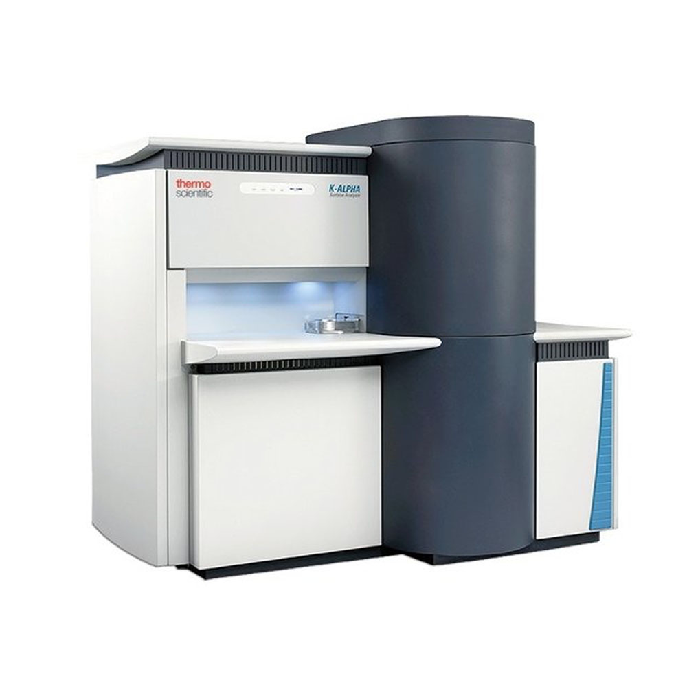
Photon sources
- Monochromated XPS - Aluminium Kα (1486.7 eV)
- Focused X-ray Spot size: 400 µm but can be as small as 30 µm with acceptable count rates, depending on the sample.
Vacuum chamber
- The vacuum in the analysis chamber can achieve as low as 1 x 10⁻⁹ mbar with several hours pumping time assuming a non- or minimally outgassing sample. The system is also equipped with the fast entry load lock.
Additional Guns
- Flood Gun - The K-Alpha is equipped with a flood gun that will ‘flood’ the analysis chamber with low energy Ar+ ions and e-.
- Ion Gun - Etching can be performed using an Ar+ ion gun. Etching can be useful for cleaning the surface of a sample and for performing a depth profile of the sample. The etching gun can be operated at 200, 500, 1000, 2000 and 3000eV with three current settings (low, medium and high).
Additional Features
- 9-sample powder holder
- Tilting module for ARXPS
High performance AFM with the following operating modes:
- Contact mode
- DART PFM
- Dual AC
- Dual AC Resonance Tracking (DART)
- Electrostatic Force Microscopy (EFM)
- Force curves
- Force Mapping Mode (force volume)
- Force modulation
- Frequency modulation
- Kelvin Probe Force Microscopy (KPFM)
- Lateral Force Mode (LFM)
- Loss tangent imaging
- Magnetic Force Microscopy (MFM)
- Nanolithography
- Nanomanipulation
- Phase imaging
- Piezoresponse Force Microscopy (PFM)
- Switching spectroscopy PFM
- Tapping mode (AC mode)
- Tapping mode with digital Q control
- Vector PFM
- Scanning Tunneling Microscopy (STM)
- Conductive AFM (CAFM)
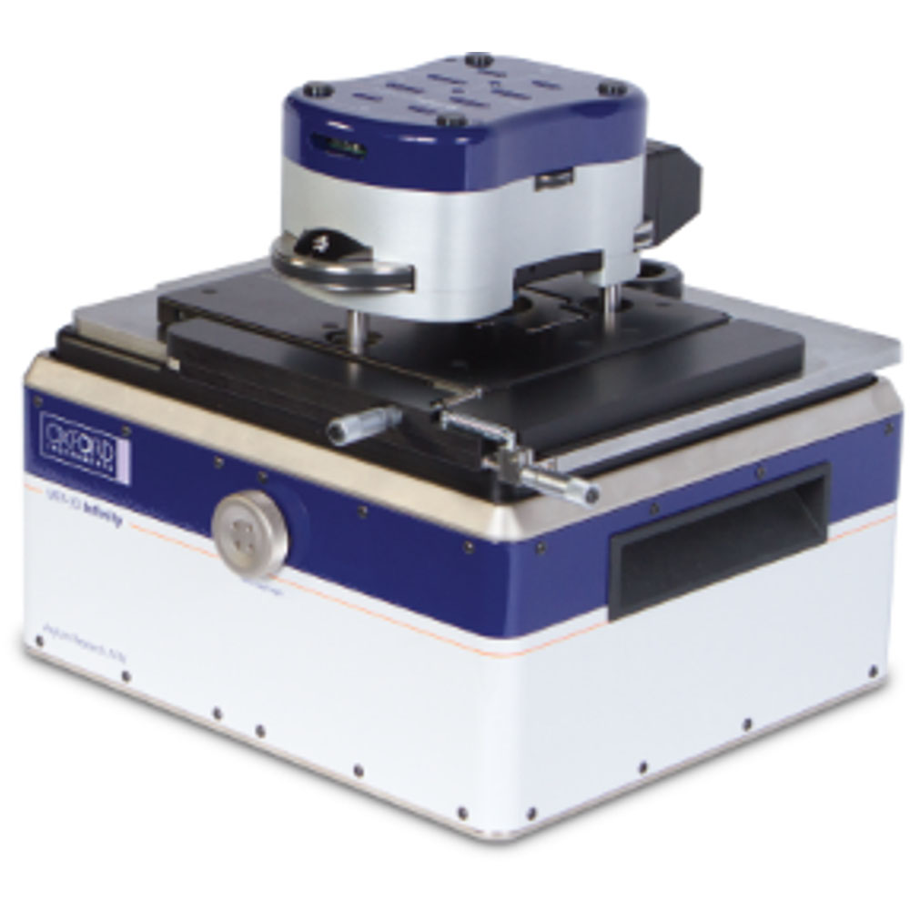
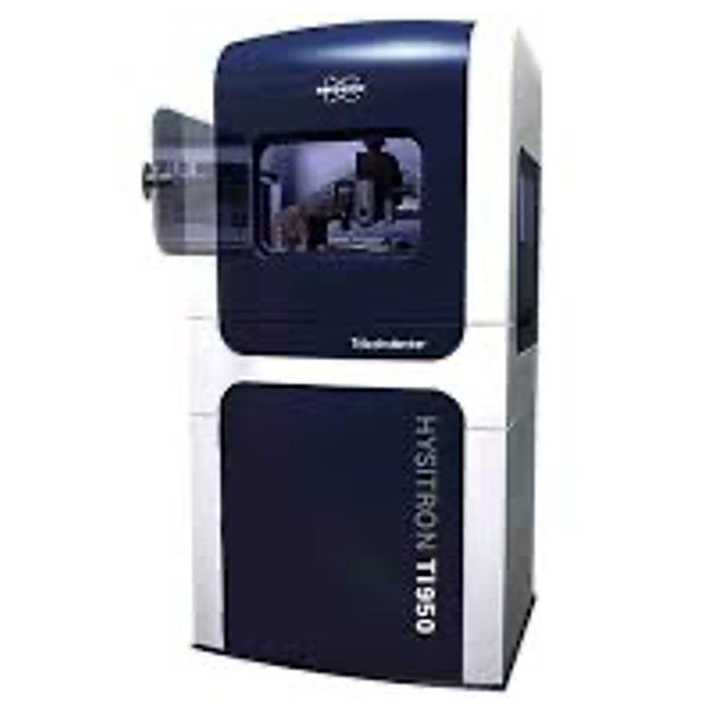
The Hysitron nanoindentation system allows for mechanical property measurements of very small areas and very thin layers. The system can also be used to map hardness as a function on position. Complex load functions can be defined changing, peak load, loading rates and load times.
- Applied loads from 100 µN to 11000 µN
Indentation probes include:
- Berkovich
- Corner cube for ultra-thin samples
- 1 µm and 5µm spear tips for soft samples
- 10µm diameter flat punch for testing nanostructures
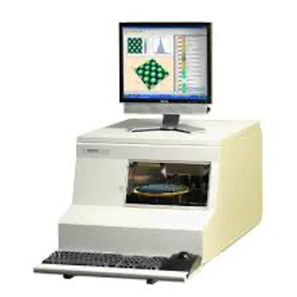
- Highly automated operation
- 2D & 3D surface profiling/imaging with lateral resolution X = 25nm, Y = 1µm
- 1mm vertical range for large topography variations
- Step height measurement with 0.6 nm repeatability
- Max. scan length >80 mm
X-Ray Equipment
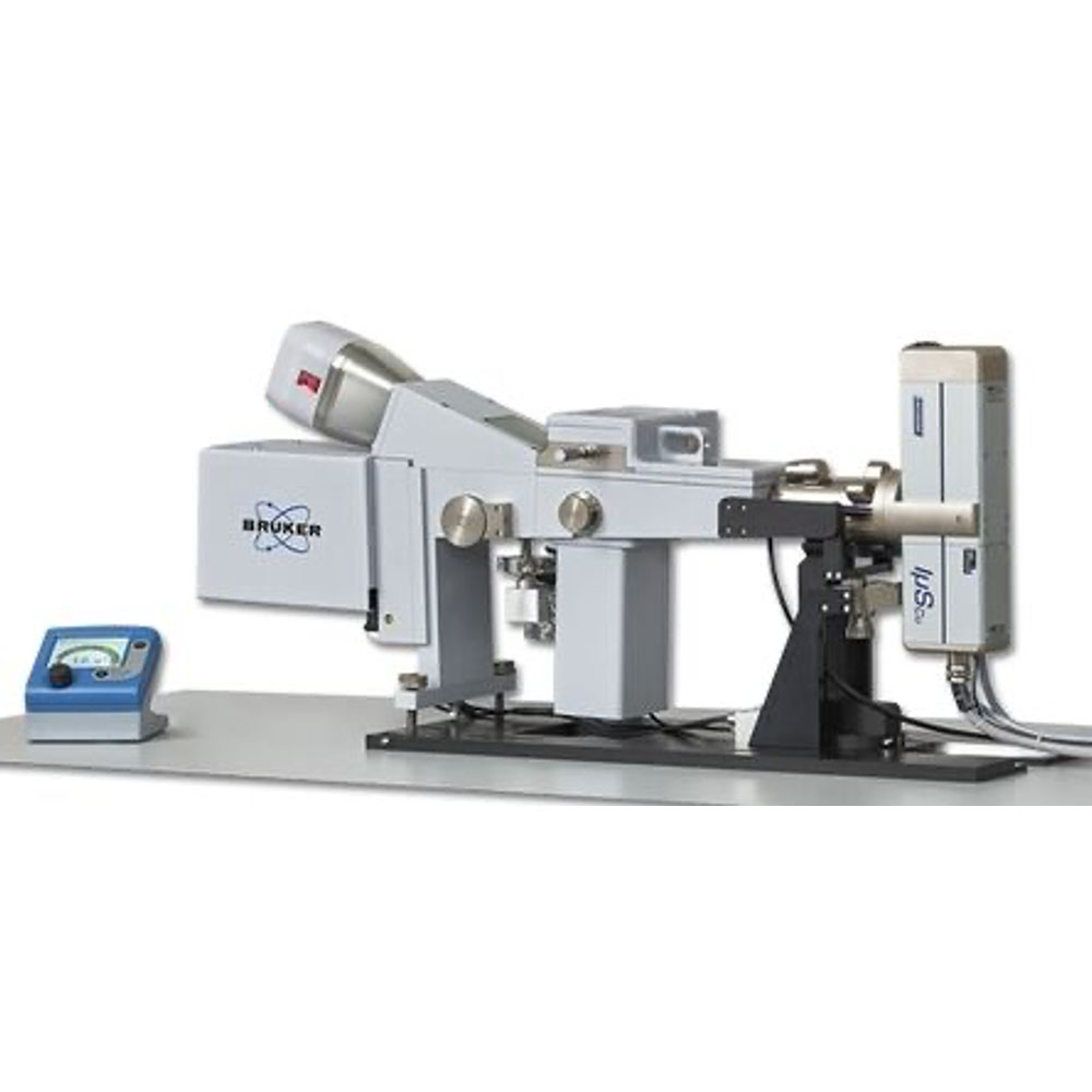
Bruker SAXS is a lab sized x-ray source for measuring the small angle x-ray scattering of large variety types materials such as powdered samples, crystalline samples, biosamples, etc.
X-ray Source:
- Bruker 1µS Copper K-α
- q-min:
- 0.004 Å-1
Small Angle Range:
- Up to 2θ = 7°, q = 0.6 Å-1
Wide Angle Range:
- 2θ = 19° - 26° q ~1.2 -1.9 Å-1
Beam Size:
- 1mm x 1mm
Sample Holders:
Basic Stage:
- Temperature range: -20°C to 120°C
- Holds any glass capillary horizontally, vacuum tight sealed.
Peltier Stage:
- Cell for pastes, powders. Flow cell for liquids and capillary spin cell for rotation.
Microcalorimeter:
- Vacuum tight quartz or glass capillaries*
- Capillary OD: 1.5mm max
- Temperature range: -30°C to 200°C
- Combined Differential Scanning Calorimetry and SAXS experiment capabilities.
- Max. Heating Rate: 5K/min
Max. Cooling Rate:
- 5K/min (200°C to 50°C)
- 1K/min (50°C to 0°C)
- 0.5K/min (0°C to -30°C)
- 20 µL (at base of capillary)
Operating Vacuum:
- 5 x 10⁻¹ Torr
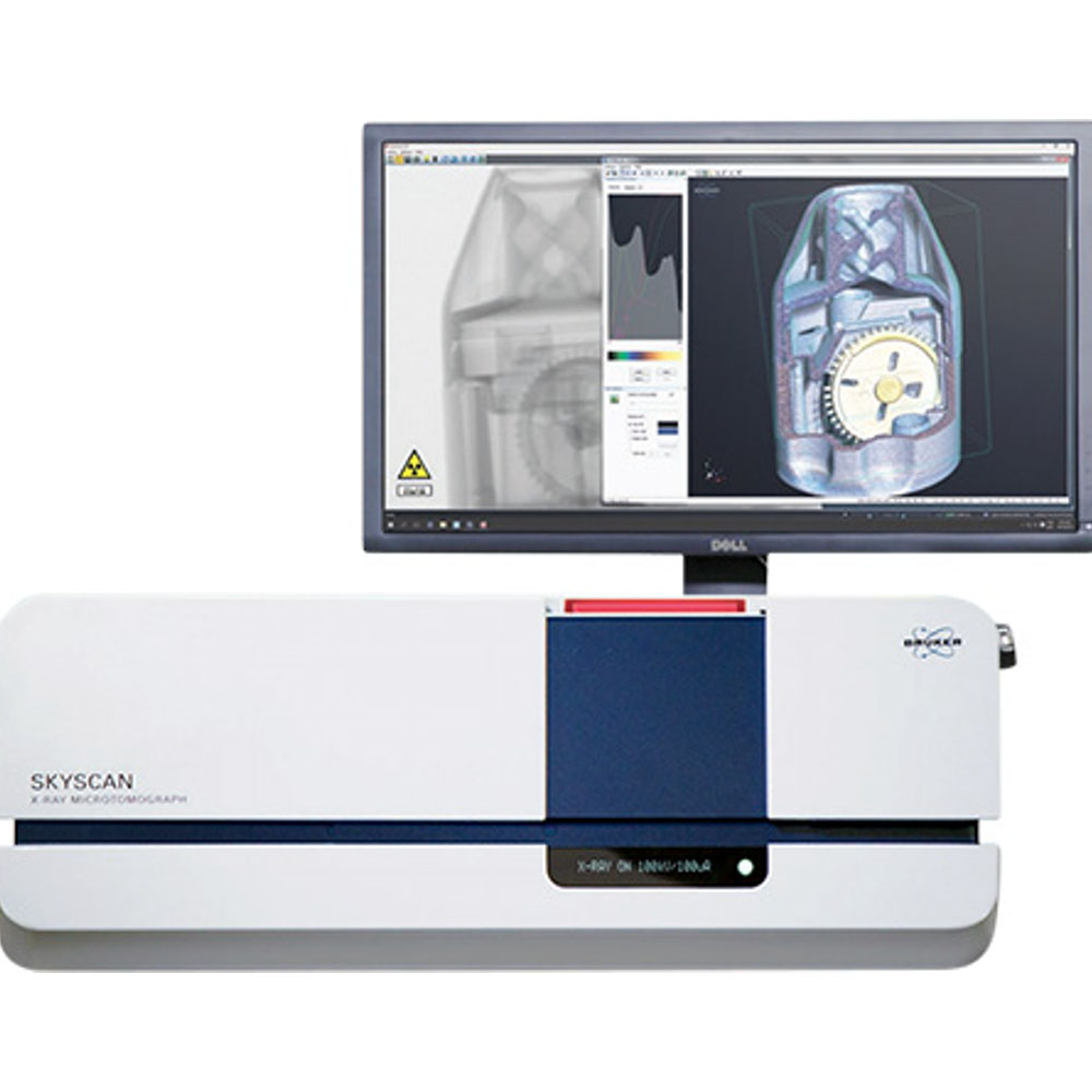
Micro-CT obtains a 3D image of the object under investigation, both from the outside and from the inside in its native state.
Features include:
- preselected sequence of actions including scanning, reconstruction and volume rendering.
- X-ray source 20-100 kV, 10 W, < 5 µm spot size @ 4 W with multiple filter options
- X-ray detector 3Mp (1944×1536 pixels) active pixel
- Maximum object size 96 mm diameter, 120 mm height
- Reconstruction GPU-accelerated FDK reconstruction as standard
- Optional stages micro-positioning, cooling, heating, compression/tension (more)
- 3D reconstruction
- software package includes programs for 2D/3D image analysis and realistic 3D visualization by surface / volume rendering of large format scanning results, data export and volume rendering on mobiles
Optical
The ALV facility is capable of a range of other experiments, including Static Light Scattering for accurate determination of particle size/distributions, Zimm plots and similar analyses for determination of Radii of Gyration, depolarized light scattering, and so on.
Typical samples include:
- Colloidal particles
- Proteins, polymers and macromolecules
- Cells, Vesicles
- Nanoparticles
- Emulsions (liquid-liquid, gas-liquid)
Particle Size:
- Typical Sizes: 10nm-1µm
Laser:
- 20mW He-Ne Laser (633nm)
Sample Preparation
General Electron Microscopy Sample Preparation
|
|
JEOL Ion Slicer |
SPI Sputter Coater |
Leica EM ACE600
|
|
|
Gatan Dry Pumping Station |
Pfeiffer Pumping Station
|
|
Polishing Equipment
|
|
Struers Labotom-3 |
Struers Secotom |
Struers Rotopol-21 |
|
|
|
|
|
Biological Sample Preparation Equipment
|
|
|
Lynx Automated Tissue processor |
Tousims critical point dryer |
Biological Sample Preparation Techniques
|
|
High pressure freezer and automated
|
Negative and positive staining
|
SEM sample preparation with CPD
|
|
|
Single and dual immunlobelling for
|
TEM and FIB-SEM tomography
|
Correlative Light and electron
|
Software
|
|
CasaXPS |
Thermo Avantage XPS |
IMOD and 3D mod |
|
|
Crystal Maker |
Digital Micrograph |
Microscopy imaging browser (MIB) |